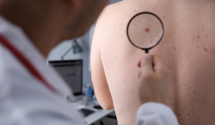
Is it a mole or melanoma? Is it harmless or not? Do I agree to treat it and how should I treat it? Should I refer? Examining our patients’ skin lesions can feel like tiptoeing through a minefield. Perhaps not unreasonably, our patients assume that as we work in medical aesthetics, we have expertise in ‘everything skin’, including moles. The problem is that most of us are not experts in skin lesion diagnosis. This raises the question: how should we approach our patient’s concerns and stay safe as medical practitioners?
We know that it takes years of specialist training in dermatology to gain the expertise required to diagnose and treat all the possible blemishes of the human skin. Luckily, many skin lesions are easy to diagnose, and you are probably already pretty good at recognising most of the ones we come across in daily practice. However, there are pitfalls, but you can improve your chances of avoiding them by sticking to a few simple rules.
In this article we will cover the presentation of common skin blemishes, with a focus on pigmented skin lesions and how to stay safe using a handful of simple safety rules for you to use in your day-to-day consultations.
What are skin lesions?
Skin lesions can arise from any organ of the skin. In medical terminology, we describe skin lesions as ‘naevus’ or ‘naevi’ in the plural form. If the naevus arises from the pigment cells (melanocytes) its correct name is a melanocytic naevus.
The medical shorthand for a melanocytic naevus is a ‘mole’. Patients tend to call everything a ‘mole’ and this colloquial meaning of the word can lead to confusion. To counter this, practitioners should refer instead to ‘skin blemishes’ or ‘skin lesions’ in your consultation before a diagnosis is made. As you examine your patient, start making diagnoses and give skin lesions their medical names as you go. Begin with the non-pigmented skin lesions you feel confident in diagnosing, such as skin tags, cherry spots, sebaceous hyperplasia, cysts, and lipomata as you are likely to be good at diagnosing all of these. DermNet NZ has a fun diagnostic tool to hone your skills and even use in the clinical setting.1 Sebaceous hyperplasia can be a little tricker to the naked eye. These are overgrowths in the sebaceous glands and tend to affect the T-zone of the face. They are small yellow-white papules with a central dip.1 When there are multiple, the diagnosis is usually clear, but isolated ones can be hard to differentiate from a basal cell carcinoma.2,3
Pigmented skin lesions
Keeping pigmented and non-pigmented skin lesions separate for yourself in this way will help reduce the likelihood of missing or inappropriately treating a melanoma. In an ideal world, all pigmented skin lesions should be assessed with the aid of a dermatoscope and the National Institute for Health and Care Excellence (NICE) recommends this for any pigmented skin lesion referred to secondary care.4 This is not practical for most non-experts or for patients themselves and naked eye examination with good lighting is adequate for the vast majority of the blemishes you will see. Keep in mind that patients with darker skin types will invariably have darker skin lesions. This can make the clinical diagnosis more difficult and, sadly, most of the literature and the references in use are still too biased towards paler skin types. This situation is partly to blame for the later diagnosis of melanoma and other skin cancers in people with darker skin types.5 There is a host of other skin lesions which can have brown pigment in them but being familiar with the listed ones will cover most scenarios. Table 1 provides tips on how to differentiate common pigmented skin lesions from each other and what they will appear like.
Freckles
Freckles are common in paler, Celtic skin types. They are usually found on the sun exposed skin of the face, upper back, chest, arms, and legs in all age groups. Simple freckles are unlikely to catch you out.7
Diagnosis |
Typical features |
Common distribution |
Freckle (ephelide) |
Multiple pale brown macules |
Face, chest, arms |
Sun/liver spot (solar lentigo) |
Well-defined uniformly coloured macules and patches |
Face, dorsum of hands |
Age spot (seborrhoeic keratosis) |
Raised warty lesions with well-defined ‘stuck on’ appearance |
Face, neck, trunk |
Dermatofibroma |
Firm papules, can be pinched |
Arms, legs |
Pigmented basal cell carcinoma (BCC) |
Shiny papules or nodules, can have crust or ulcer |
Head, neck, trunk |
Moles (melanocytic naevi) |
Pigmented macules or papules, soft to touch |
Any body site |
Melanoma |
Pigmented skin lesions with irregular features, see text |
Any body site |
Solar lentigo
A solar lentigo is the medical term for a ‘liver spot’ or a ‘sunspot’. It is the most common type of lentigo, which refers to a well-defined, flat, uniformly coloured macule or patch of skin. They are larger than freckles and are common on the face and the back of hands as we get older. Diagnosis is usually straightforward, but practitioners should be wary of isolated lentigines or ones that don’t look 100% typical. Premalignant sun damage (pigmented actinic keratosis) or melanoma in-situ (lentigo maligna) can be subtle and mimic solar lentigines.8
Seborrhoeic keratoses
Seborrhoeic keratoses arise out of lentigines as we start to feel (palpate) a raised surface or margin. We often refer to them as ‘Seb Ks’ for short. They are very common (even ubiquitous), so it’s easy to become complacent about them.9 I recommend that you consider them for what they are, a pigmented skin lesion. Proceed with care when examining likely Seb Ks and apply the safety rules outlined below. There are pitfalls here. Consider this for example: how do you pick out a melanoma in a patient with innumerable seborrhoeic keratoses?
Dermatofibromas
Dermatofibromas are small, firm pink or brown skin lesions on an extremity. They can be ‘pinched’ on examination, and you can find another similar lesion on patients who are prone to them. They are often pigmented, especially in darker skin types.9
Basal cell carcinoma
Basal cell carcinoma (BCCs) are usually raised shiny nodules, often with a crust or small ulcer (nodular BCC) or flat eczema like patches of skin (superficial BCC). They often have brown pigment within them, or they can even be heavily pigmented in people with darker skin.10 Differentiating them from an atypical seborrhoeic keratosis or melanoma with naked-eye examination can be impossible.
Moles
Moles (melanocytic naevi) come in different shapes and sizes. To keep things simple, let us think of moles in three groups:
- Flat, where the melanocyte accumulation sits at the junction between the dermis and epidermis (junctional naevi)
- A combination of flat and raised elements (compound naevi)
- Raised, where the melanocytes have started to clump together in the dermis, causing the overlying epidermis to rise and become palpable (intradermal nevus)11
Only 1% of us are born with moles.12 These are referred to as congenital naevi, and a discussion of these is outside the remit of this article. Instead, we are concerned with acquired melanocytic naevi. They accumulate throughout life but less so as we get older. Melanocytes have a general tendency to descend in the skin as we age. During this ‘maturation’, the surface of the mole tends to get paler, and the mole turns more ‘soft and wobbly’. This is a good sign and the risk of melanoma in these paler, soft, and wobbly moles is low. On the other hand, flat moles are considered of unknown potential and patients should observe them for any changes.11 Differentiating a slightly atypical mole from a superficial spreading melanoma can be tricky, even for experts using dermoscopy.
Safety rules for identifying melanoma
All of this may sound complicated or even scary. Fortunately, there are rules of thumb you can set yourself to minimise your risk of missing or treating a skin cancer inappropriately.
ABCDE rule
The first of these safety rules is the well-known ABCDE rule.13 Remember, when in doubt, consult an expert.
- ‘A’ stands for ‘asymmetry’ – can you in your mind’s eye draw a line through the mole and reflect it like a mirror image onto the other half?
- ‘B’ stands for ‘border’ – is the border of the skin lesion smooth or ragged? In other words, does the mole look like a small map of Mallorca?
- ‘C’ stands for ‘colour’ – are the colours within the mole even and symmetrical?
- ‘D’ stands for ‘diameter’ – is the lesion larger than the head of a pencil (> 6mm)?
- ‘E’ stands for ‘evolving’ – is the mole changing?
‘Ugly duckling’ rule
If you find a skin lesion you are not sure about, look for a skin lesion which looks near identical. If you find one this is clearly reassuring, since the chances of having TWO skin cancers on a single patient are clearly lower. That is not to say that you can’t have more than one skin cancer or precancerous lesion at the same time. This would normally apply more to people who have led an outdoor life, such as sportspeople or farmers, those who have lived in tropical countries, have pale skin (Fitzpatrick skin types I-II) and patients who are immunosuppressed. Fortunately, the chances of having two melanomas presenting at the same time (synchronous melanomas) are very low, even in these groups.14 The ‘ugly duckling’ rule is therefore a valid rule of thumb for many of our patients.
Beware of the isolated lesion
Any single skin lesion, however harmless looking, should get your attention. One classical pitfall is to treat an isolated verruca which later turns out to be a squamous cell carcinoma.15 Another pitfall is to be over-confident in your diagnosis of a seborrhoic keratosis. As we discussed before, seborrhoeic keratoses are best regarded as a pigmented skin lesion and, as such, the differential diagnosis is always melanoma.
Beware of the ‘new’ lesion
It is a common fallacy that most melanomas arise out of existing moles. They are more likely to come from normal skin but in the initial months or years they can mimic harmless moles.16 To catch melanomas and other skin cancers before the later stages, advise patients to be aware of any NEW skin lesions or moles.
Give it a name
This is a great rule for clinicians. If you can’t give a skin lesion a name, give yourself two options:
- Send the whole lesion or a biopsy for histology analysis (or refer to an expert)
- Bring the patient back for a follow-up in three to six months
Reassurance or treating a skin lesion without histology analysis should NOT be an option when you cannot give it a name.
Seek professional help
Diagnosing skin lesions, moles and skin cancer is clearly a skill which takes specialists years to develop. It is, however, possible to be a safe practitioner through self-directed learning, attending courses and learning from those around you who have more experience. You can contact your local NHS dermatology department and ask if shadowing sessions are available. Increasingly, dermoscopy is considered an essential skill for those who treat skin lesions.
The Primary Care Dermatology Society runs fantastic courses, if you want to gain more expertise.17 Most importantly, know your limitations and be honest with yourself and your patients if you are not sure about a skin lesion. Ask them to see their GP for an assessment and NHS referral to a dermatologist where appropriate or refer them to a dermatologist privately if they prefer. If you already treat skin lesions you must follow guidance from NICE and the British Association of Dermatologists.4,18
Take skin lesions seriously
Consider your learning needs in the context of your practice and use the resources out there to address them. Use the simple safety rules we have outlined, know your own limitations, and refer to experts when in doubt. This way you are unlikely to contribute to harm for your patient by false reassurance or, worse, treating a skin lesion which later turns out to be cancerous.
References:
1. DermNet NZ, ‘DermDiag’, <https://dermnetnz.org/dermdiag>
2. Skin Cancer Foundation, ‘Basal Cell Carcinoma Overview’, 2022, <https://www.skincancer.org/skin-cancer-information/basal-cell-carcinoma/>
3. Zaballos P, et al., ‘Dermascopy of Sebaceous Hyperplasia’, Archives of Dermatological Research, 2005, <https://jamanetwork.com/journals/jamadermatology/article-abstract/395657>
4. National Institute for Health and Care Excellence, ‘Melanoma: assessment and management NICE guidelines (NG14), 2015, <https://www.nice.org.uk/guidance/ng14>
5. Ramji R, et al., ‘Melanoma in skin of colour’, DermaNet NZ, 2017, < https://dermnetnz.org/topics/melanoma-in-skin-of-colour>
6. Mowlabaccus W, et al., ‘Common benign skin lesions’, DermaNet NZ, 2020,
<https://dermnetnz.org/topics/benign-skin-lesions>
7. Griffiths C, et al., ‘Rook’s Textbook of Dermatology’, ninth edition, 2016.
8. Chan B, ‘Solar Lentigo’, DermNet NZ, 2014, < https://dermnetnz.org/topics/solar-lentigo>
9. Tanaka M, et al., ‘Key Points in dermatoscopic differentiation between lentigo maligna and solar lentigo’, Journal of Dermaology, 2011, p.53-8.
10. Nasr I, et al, ‘British Association of Dermatologists guidelines for the management of adults with basal cell carcinoma 2021’, Wiley Online Library, 2021, <https://onlinelibrary.wiley.com/doi/10.1111/bjd.20524>
11. McCalmont T, ‘Melanocytic Nevi’, Medscape, 2019, <https://emedicine.medscape.com/article/1058445-overview>
12. Jacobs A, Walton R, ‘The incidence of birthmarks in the neonate’, Pediatrics, 1976, p.218-22.
13. Cancer Research UK, ‘Symptoms’, 2020, https://www.cancerresearchuk.org/about-cancer/melanoma/symptoms
14. Claeson M, et al., ‘Multiple Primary Melanomas: A Common Occurrence in Western Sweden’, Advances in Dermatology and Venereology, 2016,<https://www.medicaljournals.se/acta/content/html/10.2340/00015555-2598>
15. Keohane SG, et al, ‘British Association of Dermatologists guidelines for the management of people with cutaneous squamous cell carcinoma 2020’, Wiley Online Library, 2020, <https://onlinelibrary.wiley.com/doi/full/10.1111/bjd.19621>
16. Cymerman R, et al., ‘De Nova vs Nevus – Associated Melanomas: Differences in Associations with Prognostic Indicators and Survival’, Journal of the National Cancer Institute, 2016, <https://pubmed.ncbi.nlm.nih.gov/27235387/>
17. Primary Care Dermatology Society, ‘General Dermaology Events’, <https://www.pcds.org.uk/events/general-dermatology>
18. British Association of Dermatologists, ‘NICE Diagnostic Assessment Programme: Vivascope 1500 and 3000 for detecting and monitoring skin lesions’, 2015, https://www.bad.org.uk/ResourceListing.aspx?sitesectionid=1139&itemid=7111