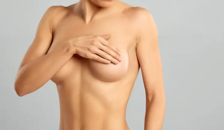
Search

Lifting the Breast with Radiofrequency

The breasts are a uniquely important part of the female body, with both physiological and psychosocial roles. In many works of art throughout history, breasts have been portrayed prominently, and with many symbolic associations, most frequently with fertility.

Views of their importance and acceptance have metamorphosed through time and have reflected upon the views of women of the period and society in general. It therefore comes as no surprise that aesthetic surgery of the breast is one of the most common procedures performed by plastic surgeons today, as a proportionately developed breast continues to be an important feminine feature.1

Breast ptosis

Breast droop, or ptosis, is an extremely common occurrence, following weight loss, pregnancy, breastfeeding, and more generally as a consequence of normal ageing. The result is a loss of youthful shape and contour, frequently associated with a change in the position and orientation of the nipple and areola complex.2 This results in aesthetic detriment and deviation from the broadly accepted ‘ideal’ breast shape,3 described as a 45:55 ratio between the upper and lower pole of the breast, with an upward pointing nipple at an angle between 10-20 degrees, and with a straight or gently concave upper slope and a convex lower slope.2

Breast ptosis can be classified using the Regnault classification system, which describes the degree of droop by the relative position of the nipple areola complex and the inframammary fold (Figure 1).4

The pathophysiology of breast ptosis centres around the effect of gravity, parenchymal loss, loss of elasticity, decreased integrity of fascial support and of Cooper’s ligaments, and the thinning and stretching out of skin. As a result, breast position is lower on the chest wall, breast contour changes, and upper pole fullness is lost.2

Breast restoration with radiofrequency

Mastopexy is a surgical procedure that seeks to restore a more youthful shape and contour of the breast, restoring texture, repositioning the nipple areola complex within the breast mound, and tightening the skin envelope. Traditionally this can be achieved through a variety of surgical approaches, which vary in indications depending on breast features, but all share some common drawbacks. Scarring and the need for general anaesthetic are frequent reasons why women who have experienced ptosis frequently choose to defer or opt out from addressing their cosmetic concern.

It is in this context that non-surgical interventions may be considered. In our clinic, we use radiofrequency assisted lipocoagulation (RFAL) combined with radiofrequency (RF) microneedling. This is a ‘scarless’ procedure that can be performed under tumescent local anaesthetic in the clinic – with minimal risks and post-procedural downtime. In the appropriate patient, it is capable of producing a desirable degree of lift and contouring of the breast without any of the downsides of a surgical mastopexy. The practitioner performing the procedure should be a General Medical Council registered doctor with full licence to practice. The use of the device requires CQC registration.

RF is a form of electromagnetic current that can be delivered to a variety of tissue types to generate thermal energy.5 When RF is applied to skin and underlying soft tissue, it generates contraction through two mechanisms. The first is shortening and thickening of collagen fibres and the second is initiation of a wound-healing cascade to trigger neoangiogenesis, neocollagenesis, and elastin reorganisation over the following three to four months.6 This explains why some results are noticed early on following the procedure but, in our experience, the more dramatic results manifest at three to six months post procedure.

In our practice, we use BreastTite, which is part of the BodyTite platform. This is a bipolar device where electromagnetic energy is transmitted from a generator to the tissue between the two electrodes of a directional handpiece (Figure 2). It has controlled energy, which flows between the tips of internal and external sensors. We believe it’s important to use a device that has real-time internal and external temperature monitoring. This is so that when a pre-set temperature is reached, energy delivery stops so it does not overheat and cause injury to skin and surrounding tissue.7

Consultation

As is the case with all cosmetic and aesthetic work, the pre-procedural consultation is essential to assess the patient, determine their needs and expectations, and select the most appropriate approach. It is essential to understand the patient’s specific concerns about their breasts, and to foster discussion and realistic expectations. Goals with regards to shape, size and fullness must be established. It is essential to manage these and to be transparent with regards to the potential of the technique. Whilst RFAL will likely produce a degree of tissue tightening and contouring for the majority of patients, it would be an inadequate choice for patients with grade 3 ptosis or with moderately large or large breasts. These patients are better candidates for surgery.

Furthermore, patients seeking volume increase, particularly at the upper pole of the breast, will be unlikely to be satisfied with the results of RFAL, and should instead be counselled towards augmentation mastopexy and referred onwards as appropriate.

The ideal patient for this procedure is someone with grade 1 or grade 2 ptosis (Figure 1), with small to moderately-sized breasts, typically not larger than C cup. In our experience, satisfactory results with noticeable contouring can be achieved in larger breasts also, but these should constitute the exception rather than the rule.

At the time of the initial consultation, it is essential to assess breast health, and ensure that a full history with regards to previous or current breast symptoms and family history of breast disease is determined. A standard breast examination should be routinely carried out and any symptoms or findings should be investigated as per clinical guidelines prior to committing the patient to the procedure.

In our practice, women aged 40 and above – even if asymptomatic and with no significant personal or family history – are referred for mammography prior to being considered for RFAL. Risks, such as pain, swelling, bruising, seroma, burns, scarring (at the entry port), failure to achieve lift or development of asymmetry, and benefits are discussed with the patient. Patients with a history of breast cancer, diabetes, poorly controlled cardiovascular disease, coagulopathy, steroid use for the management of autoimmune conditions, and smokers, are considered not suitable for this procedure and should generally be discouraged from pursuing cosmetic surgery of the breast in view of the increased risk of complications.

Standard measurements are taken with particular attention to the sternal notch-to-nipple distance, nipple-to-inframammary fold distance, breast base and breast height on both sides (Figure 3), and clinical photography is taken to document breast position and shape.

Treatment protocol

The procedure is ideally performed under tumescent local anaesthesia and excellent levels of pain control allow the procedure to be safely performed in combination with 5mg diazepam, 30mg dyhidrocodeine and 1g paracetamol orally 30 minutes prior to the beginning of the procedure. This method of local anaesthetic is commonly employed in liposuction,8 and creates adequate space in the relatively avascular fascial layer of the breast separating the skin from the breast gland, to allow the internal cannula of the device to travel, and optimises conductivity for the RF between the internal and external electrode.8 This avoids bad coupling and reduces the risk of thermal injury. In our practice, tumescent anaesthesia is established infiltrating approximately 125ml of an isotonic solution of normal saline containing 10ml of 2% Xylocaine with 1:200,000 adrenaline for each breast. This results in a well-tolerated procedure with a short recovery phase and ideal safety profile.

Standard clinical photography and paperwork are carried out, ensuring informed consent. The patient is placed in the supine position on the table, the skin is prepared with chlorhexidine solution and the field is draped with sterile drapes.

Small volumes of 2% Xylocaine are administered at the chosen entry points, typically:

  1. At the border of the areola at the 12 o’clock position
  2. Towards the axillary tail of the breast
  3. Medially along the inframammary fold
  4. At the lateral aspect of the breast just above the inframammary fold

Entry ports are created with the tip of an 18 gauge needle, and then extended to 2-3mm in size using the tip of a size 11 scalpel blade.

The tumescent anaesthetic solution is infiltrated using a cannula and across the entire surface of the breast (sparing the nipple and areola complex) to the circumferential edge of the breast footprint.

The device is set up and prepared by an assistant, and the handpiece is connected. Standard settings include external temperature cut-off at 38°C, internal cut-off at 60°C, treatment time of 120 seconds.

Following infiltration of the tumescent, the internal cannula of the handpiece is introduced through the periareolar port and allowed to glide to the edge of the breast. Lubricating gel is applied to the skin surface over the area to be treated to improve conductivity. Energy is then delivered on slow withdrawal to allow the heating of the treated tissue to the desired temperature cut-off (Figure 4).

Typically, energy is delivered one breast quadrant at a time, to allow even distribution of treatment. A total of 10-15kJ are delivered to each breast, with some variability between cases dictated by breast size and degree of correction required. The contralateral breast is then treated following the same principles.

At the end of the procedure, following further skin preparation, we will treat both breasts with a RF microneedling handpiece (we use Morpheus8), over the entire surface of the breast, at 3mm of depth, with energy setting 45 and overlap of 50% (Figure 5).

Entry ports are closed with either a single 5/0 nylon suture, or with Steri-Strips. The wounds are then protected with padded waterproof dressings.

The whole procedure takes two-to-three hours and is only carried out once. The patient is discharged with a one-week course of prophylactic antibiotic and regular analgesia. Follow-up appointments are scheduled at one week (to remove sutures if any were placed) then one, three and six months to assess the development of post-procedure results.

Post treatment

In our experience, derived from management of post-surgical breast patients, it is essential that the patient is instructed to wear a supportive post-surgical bra 24 hours a day for four weeks, and then during the daytime for a further four weeks. Patients are allowed to return to normal activity from the following day, but are advised to avoid particularly strenuous exercise and sporting activity for two to four weeks.

The expectation from the procedure is that at the time of the three-month review, noticeable results in terms of lift and improved contouring will have begun to consolidate.

All patients treated describe the procedure as having rejuvenated and firmed their breasts, with the majority having noticed a good degree of lift, an improvement in breast shape and a noticeable improvement in skin quality, with reduction in pores and improvement in skin tone.

In our experience, assuming adequate patient selection, RFAL can achieve between 1.5cm and 3.5cm of lift when measured as nipple-areola complex repositioning (Figures 6 & 7). Furthermore, improved contouring with a rounder fuller appearance is achieved in all patients we have treated. The technique is also adept at correcting mild to moderate asymmetry in breast position and results are expected to last five years (Figure 8).

Conclusion

Breast ptosis is one of the most common concerns women experience from an aesthetic standpoint. Whilst surgical mastopexy is an extremely effective way of addressing this concern, many patients defer or rule out engaging with it because of their concerns particularly surrounding general anaesthetic and conspicuous scarring.

RFAL is a minimally invasive, non-surgical, virtually scarless approach that can be safely and comfortably carried out under local anaesthetic in the outpatient setting with minimal risks.

It is ideal for patients with small-to-moderately sized breasts and grade 1 or 2 ptosis, for whom it consistently achieves improved contour as well as a 1.5-3.5cm lift in nipple areola complex position, with satisfactory enhancement of breast cosmesis without the drawbacks of surgery.

References:

1. Khan UD. An overview of aesthetic surgery of the breast. Plast Aesthet Res 2016;3:1-2.

2. Rinker B, Veneracion M, Walsh CP. Breast ptosis: causes and cure. Ann Plast Surg. 2010 May; 64 (5):579-84.

3. Mallucci P, Branford OA. Concepts in aesthetic breast dimensions: analysis of the ideal breast. J Plast Reconstr Aesthet Surg. 2012 Jan; 65(1):8-16.

4. Regnault P. Breast ptosis. Definition and treatment. Clin Plast Surg. 1976; 3:193-203.

5. Greene RM, Greene JB. Skin tightening technologies. Facial Plast Surg. 2014 Feb; 30(1): 62-67.

6. Zelickson BD et al. Histological and ultrastructural evaluation of the effects of a radiofrequency-based non-ablative dermal remodelling device. Arch Dermatol. 2004 Feb; 140(2): 204-209.

7. Theodorou S et al. Soft tissue contraction in body contouring with radiofrequency-assisted liposuction: a treatment gap solution. Aesthet Surg J. 2018 Jun; 38(2): S74-S83.

8. Klein JA. Tumescent technique for local anesthesia improves safety in large-volume liposuction. Plast Reconstr Surg. 1993; 92(6):1085–1098.

Share this article: