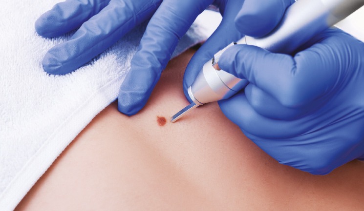
Before you start treating skin blemishes, practitioners need to hone their lesion recognition skills. This is because they must minimise the risk of inappropriately removing or destroying a lesion which turns out to be cancerous.1 Incorrectly treating a blemish could lead to potential harm for the patient.
In this article, I will consider how to avoid this, and outline common pitfalls in skin surgery.
Preparation
Skin surgery is a regulated activity in the UK, so the premises must be registered and inspected by the relevant regulator in your country or region of the UK (CQC, CIW, HIS, RQIA). It will likely take a few months to set up a new skin surgery service to meet the regulatory demands and ensure that you have the appropriate equipment and safety protocols in place. Practitioners must have completed an approved training course in skin surgery which offers a certificate of completion, and have medical indemnity before starting.2
Infection control
Practitioners will already be familiar with appropriate infection control measures. Infection risk is higher in skin surgery than in aesthetics, so practitioners should pay attention to hand hygiene, choice of gloves, skin preparation, drapes, use of aprons and hats, as well as careful aseptic technique. Practitioners need written standard operating procedures for each element of the service and must train staff accordingly. This includes a written protocol on hand washing, opening a surgical pack, clearing up post-treatment, instrument counting and suture removal. Instruments need counting after surgery to minimise the risk of the patient leaving the premises with a needle or sharp object on their clothes or hair, leading to injury to them or others they meet.3 Practitioners must have a separate needle-stick policy and procedure in place.3
Consent
As part of the Good Medical Practice guidelines, practitioners must obtain signed consent from patients, with details of the procedure, body site treated, side effects and possible complications. You also should assess the patient’s ability to give informed consent.4 The regulator will expect practitioners to have a consent and capacity policy and procedure detailing how capacity is assessed and what to do when capacity is in question.5
Local anaesthetic
Many skin surgeons use 1-2% lidocaine (10-20mg/ml) mixed with adrenaline 1:80,000-1:200,000. This works well and is freely available to medical practitioners from suppliers. The adrenaline causes vasoconstriction, helping to reduce bleeding.6
How are skin lesions removed?
Cautery
Cautery is the process of delivering electricity to the skin, causing heat and a controlled burn. Cautery machines have energy settings according to the area being treated. Many practitioners use a Hyfrecator device, which is easy to use and has a good safety record.7 Other reputable manufactures include Schuco. To cauterise a lesion, touch the lesion until it chars black. Wipe the blemish away with a swab and repeat until the lesion is gone and bleeding has stopped. Generally, two to three cycles are sufficient. Cautery on its own is suitable to treat spider naevi, small skin tags, cherry haemangiomas, syringoma, milia and sebaceous hyperplasia.3,8
Snip and cautery
In my view, this is the best way to remove larger skin tags as they are easy to grab with a pair of forceps, allowing the practitioner to snip the base with surgical scissors. Place a small amount of anaesthetic at the base and cauterise to stop bleeding. Occasionally, this technique may result in a full thickness wound. If this happens and the bleeding does not stop with cautery, consider placing one to two haemostatic sutures on the skin and remove a week later.3,8
Curettage and cautery
I use a 4 or 7mm ring curette according to lesion size. After numbing the area, scrape from the periphery towards the centre to remove tissue and cauterise to stop bleeding. Two to three cycles of scraping and cauterising may be required before the lesion is removed.3,8
In my experience, this technique works for superficial lesions such as seborrhoeic keratoses. It is also useful when treating larger cherry haemangiomas, syringoma, milia, sebaceous hyperplasia or removing remnants of lesions after snipping or shave excision. Curettage and cautery is a common treatment for low-risk skin cancers or precancer, such as actinic keratosis, Bowen’s disease and superficial basal cell carcinoma.9
When treating suspected skin cancer, you must follow guidelines from the National Institute for Health and Care Excellence (NICE).10 If it isn’t within your scope of practice to treat skin cancers, then refer your patient to a suitable practitioner who has experience in treating lesions.
Shave excision
Shave excision is where a lesion is removed by a blade or surgical knife, without breaching the deeper dermis or requiring sutures to repair the wound. With this technique you rock the device gently as you advance it under the lesion, heading down until reaching the centre of the lesion, then up and out the other side. If the lesion isn’t removed at once, practitioners may use a curette to finish the procedure and cauterise to stop bleeding. This is a preferred technique for removing exophytic lesions, such as dermal naevi and larger seborrhoeic keratoses.3,8
Punch biopsy
This is a small ‘apple-corer’, used in the diagnosis of a lesion or rash. They can be used to remove small skin blemishes, such as melanocytic naevi. After placing the anaesthetic, push the tool against the skin while twisting it until the dermis is pierced. Use non-toothed forceps to lift the sample up and snip through the subcutaneous fat with surgical scissors. Practitioners should remove some fat to ensure full thickness removal and a sample for analysis. Interrupted stitches should be placed on the area to stop bleeding from a small wound as well as closing the defect. Stitches can be removed after seven days on the face and 12-14 days on the body.3,8
Surgical excision
Surgical excision refers to the removal of a lesion by cutting around it through the skin and the tissue underneath the blemish. It is the appropriate way to remove larger and deeper lesions, such as moles, cysts and suspected or confirmed skin cancers. Practitioners should mark out the lesion and a margin of healthy skin and then an ellipse (‘boat shape’) where the length of the excision is three times the size of the lesion. This should result in a flat scar.3,8
After the skin is numbed, use a surgical blade to incise through the dermis and use forceps and scissors to cut through the subcutaneous fat until the sample is free. Cautery is usually sufficient to stop bleeding, but practitioners should know how to clamp and tie off a bleeding vessel. Once the wound is dry, deep dissolvable stitches are placed to oppose the edges, followed by interrupted non-dissolvable stitches to the surface. Sutures can be removed after seven days on the face and 12-14 days on the body.3,8 An important tip is to ensure no flammable fluids are used to sterilise the skin. This can risk causing a fire or burn to the patient’s skin during cautery. I would remove hazardous materials from your surgical suite. Safe options include 0.05% aqueous chlorhexidine (Unisept) or Clinisept+ Skin, with other brands available.
High-risk sites
If practitioners are cutting through the skin, they need to know about high-risk areas. There are locations on the body where motor nerves run close to the skin. Injury to these will cause permanent weakness of the muscles served by the nerve. Examples include injury to the facial nerve over the zygoma, resulting in a weak frontalis muscle and brow drop, and injury to the accessory nerve in the lateral neck, resulting in a weak shoulder. Injury to a blood vessel not responding to cautery can be repaired by clamping the vessel and tying it with a suture. If bleeding is controlled, the patient will not come to any harm. These high-risk sites will be covered in detail on a competent training course in skin surgery.3,8
Pitfalls
As with any area of medical practice, there are pitfalls in lesion treatment. To help avoid these issues, follow the advice given in the boxout.
Wrong diagnosis
Could a melanoma be ‘hiding’ amongst your patients’ solar lentigines, seborrhoeic keratoses and moles?1 Before treatment, ask yourself the questions in the box above.
Wrong technique
The most common ‘mistake’ is to carry out a shave excision of a ‘mole’, which turns out to be a melanoma. A shave excision may not get to the base of a melanoma, meaning that it cannot be ‘staged’ for treatment planning and prognosis. The way to remove any lesion which could be a melanoma is by complete excision with 2mm margin of healthy-looking skin.9
Recurrence
A skin lesion may recur if it is incompletely removed. Incomplete removal of a skin cancer is detrimental for your patient, and therefore practitioners should be careful about treating a lesion without histological analysis (see steps in the boxout).
Rare entities
All organs of the skin can turn cancerous, and the resulting tumours can initially look innocuous, such as Merkel cell carcinoma. Another rare entity is the seborrheic keratosis-like melanoma – an almost impossible diagnostic challenge. It’s vital that practitioners consider the possibility of rare entities and refer to a specialist if they cannot treat themselves.
Aftercare
For small wounds, a spot plaster will suffice, however for larger wounds use a breathable, absorbent dressing. Advise patients to change it every day from day two to three onwards, or use petroleum jelly or a antimicrobial product if they wish to leave the area undressed. The wound should not be soaked with water but cleaned gently every day. Patients should avoid physical activity for a few days or until stitches are removed. Encourage patients to contact the clinic if they have concerns, so any complications can be treated early.8
Complications
Infection occurs in up to 10% of skin surgery procedures.11 The rate is low for procedures where no stitches are used, but highest for large excisions and in skin cancer surgery.11 Signs include redness, pain, swelling and oozing of pus. These symptoms are common in the first two to three days, and practitioners may reassure the patient if things are improving over time. However, if symptoms are worsening, review the patient in-clinic.8
When treating lesions, blood can be expected. If the wound oozes blood, advise the patient to press on the area for 20 minutes. If this doesn’t work, additional haemostatic sutures may be required.8 A fluctuant area with an overlying bruise suggests collection of blood in the wound. Advise the patient to cool the area, rest and use painkillers. Anti-inflammatory drugs can aggravate bleeding and haematomas. Rarely, a haematoma may need lancing.8
Dehiscence (opening up of a stitched wound) is a sign of poor suture technique, infection, patient factors such as smoking or diabetes or not complying with aftercare instructions.12 Practitioners should dress the wound every two to three days and wait for it to heal from the bottom and sides. Skin lesion treatments may result in scar formation. A hypertrophic scar is a thick scar corresponding to the footprint of the procedure, whereas a keloid scar grows outside the confines of the procedure. The risk is highest in areas of skin tension, particularly the upper chest, back and shoulders, and is more common in Fitzpatrick skin types IV-VI.8 Treatment options include silicone gel, topical steroid under occlusion, steroid injections and laser. None are satisfactory and even when treatment is successful, the cosmetic outcome tends to be poor.8
Safely treat skin lesions
Skin surgery is a fulfilling area of practice with high patient satisfaction.13 There are many training providers which offer courses in lesion diagnosis, dermoscopy and skin surgery. Being able to recognise common lesions and offer treatments is positive for your patients and may fit well into your practice. There are pitfalls, however, and you must ensure that you follow guidance by NICE and fulfil the requirements laid down by the medical regulator of the nation where you practice.
References:
1. Thorsteinsson K, ‘Diagnosing Skin Lesions’, 2022, <https://aestheticsjournal.com/feature/diagnosing-skin-lesions>
2. NHS, ‘Quality Standards for Dermatology’, 2022, <https://www.pcds.org.uk/files/gallery/dermatolog-standards-final.pdf>
3. British Society for Dermatological Surgery, 3’BSDS/BAD Guidelines’, 2011, <https://bsds.org.uk/resources/bsds-bad-guidelines/>
4. General Medical Council, ‘Good medical practice’, 2014, <https://www.gmc-uk.org/ethical-guidance/ethical-guidance-for-doctors/good-medical-practice>
5. Care Quality Commission, ‘Regulation 11: Need for consent’, 2022, <https://www.cqc.org.uk/guidance-providers/regulationsenforcement/ regulation-11-need-consent>
6. Niemi G, ‘Advantages and disadvantages of adrenaline in regional anaesthesia’, Best Practice and Research in Clinical Anaesthesiology, 2005, p.229-45.
7. Weyer C, et al., ‘Investigation of hyfrecators and their in vitro interference with implantable cardiac devices’, Dermatologic Surgery, 2012, p.1843-8.
8. Cunliffe T, Chou C, ‘Primary Care Dermatology Society – Skin Surgery Guidelines’, 2007, <https://www.pcds.org.uk/files/gallery/skin_surgery_guidelines.pdf>
9. Mazzoni D, Muir J, ‘A guide to curettage and cautery in the management of skin lesions’, Australian Journal of General Practice, 2021, p.893-897.
10. National Institute for Health and Clinical Excellence, ‘Improving outcomes for people with skin tumours including melanoma(update)’, 2010, <https://www.nice.org.uk/guidance/csg8/evidence/2010-update-the-management-of-lowrisk-basal-cellcarcinomas-in-the-community-updated-recommendations-andevidence-on-this-topic-only-pdf-7022614429>
11. Dixon A, et al., ‘Prospective study of wound infections in dermatologic surgery in the absence of prophylactic antibiotics’, Dermatologic Surgery, 2006, p.819-26.
12. National Library of Medicine, ‘Wound Dehiscence’, 2022, <https://www.ncbi.nlm.nih.gov/books/NBK551712/>
13. JAMA Surgery, ‘Patient-Reported Outcomes and Factors Associated with Patient Satisfaction After Surgical Treatment of Facial Non-Melanoma Skin Cancer’, 2018, p.179-181.