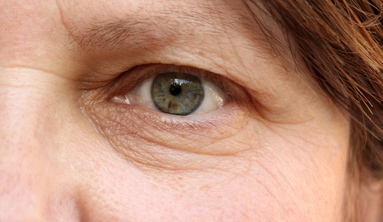
Blepharoplasty is one of the most commonly performed cosmetic operations.1 The eyes are an important aesthetic facial feature and eyelid skin is one of the first areas of the face to show signs of ageing.2 By understanding the anatomy and associated facial changes over time, non-surgical and surgical treatment options can be considered. This article describes the ageing process around the eye and how to examine this area in the context of blepharoplasty, and the different blepharoplasty methods.
Ageing process in the periocular region
The eyelid is made up of several layers. From superficial to deep; the skin, subcutaneous tissue, orbicularis oculi muscle, orbital septum and tarsal plates, smooth muscle and the conjunctiva. The upper eyelid contains the insertion of the levator palpebrae superioris muscle, which forms the upper eyelid skin crease (Figure 1).3
The intricate eyelid structure results in complex periocular changes over time. One of the first signs is the presence of excess skin, termed dermatochalasis. With ageing, the skin becomes loose and inelastic as collagen production declines and this is accelerated by UV exposure. Excess skin can be so severe that it can start to obstruct vision in the superior visual field. If the orbital septum and ligaments weaken, the support of fat and muscle is reduced.4,5
In the upper eyelid, there are two fat pads which commonly prolapse forward, namely medial and central posterior with respect to the orbital septum. In the lower lid, there are three fat pads, medial, central and lateral (Figure 2). As facial tissues descend and weaken there is an associated cheek ptosis which deepens the nasojugal fold, called the tear trough deformity. The levator aponeurosis can stretch resulting in ptosis of the upper eyelid. The smooth muscle retractors in the lower lid can also weaken, resulting in an entropion or ectropion. These age-related changes underpin the concerns of many patients seeking rejuvenation of the periocular area.4,5

Periocular evaluation
Assessment starts with a thorough history focusing on medical, ophthalmic and aesthetic aspects. A mirror is very useful at this stage. It is important to understand the patient’s concerns and manage their expectations realistically. I always offer surgical patients an optional complementary second consultation to discuss any outstanding issues. I also send a detailed medical report and information on aftercare. Practitioners should review the brow position; a ruler can be used to look for brow asymmetry and any brow ptosis should be noted and addressed prior to surgical or non-surgical blepharoplasty.6
Brow position does not change in patients who undergo upper eyelid blepharoplasty for simple dermatochalasis.7 A retrospective study by Goldberg et al. found the mean preoperative brow position to be approximately 17.5mm above the pupil, with the eye in primary position.7 For your assessment, the upper eyelid position should be inspected and any asymmetry, excess skin or fat pad prolapse should be noted. The measurements start with the palpebral aperture, which is the distance between the upper and lower eyelid; it usually measures 10mm.8

Next, to aid the diagnosis of a ptosis, the distance between the pupillary light reflex and the upper eyelid should be measured, which is called the marginal reflex distance one (MRD1), depicted inFigure 3 and described in Figure 4.8 If a ptotic eyelid is ignored during blepharoplasty assessment the patient will likely be disappointed with the outcome because the upper eyelid will still be low. The upper eyelid skin crease should also be assessed. It is thought to be formed by the insertion of levator muscle fibres and is approximately 8-10mm in women and 7-8mm in men.8
The lower lid should be observed for skin quality, excess skin and fat pad prolapse. The presence of horizontal laxity can be assessed by pulling the lid laterally in both directions, which will measure the strength of the medial and lateral canthal tendons. Other measurements are usually taken at this point such as the presence of inferior scleral show, the distance between the inferior corneal limbus and lower eyelid and the distance from the pupil to the lower eyelid – also known as MRD2 (Figure 4).

Non-surgical blepharoplasty
Non-surgical blepharoplasty is an umbrella term which compromises various treatment options to target excess periocular skin.
Resurfacing treatments
The most basic options are periocular chemical peels and microneedling.10 Chemical peels can be used to treat fine lines and dark circles or used in combination with a transconjunctival blepharoplasty.11 One must be careful with the chemical concentration as the skin thickness may be as little as 0.2mm in this area.12
Products that can be titrated to produce superficial to moderate-depth peels can include glycolic acid and trichloroacetic acid.13 Practitioners may also use laser resurfacing treatments such carbon dioxide (CO2) and Er:YAG lasers with good results in the appropriate patient, however it is not able to address fat prolapse or volume loss.14
The CO2 ablative fractionated laser vaporises the epidermis and reticular dermis through thermal energy, thus causing deep tissue tightening.15 The Er:YAG laser tightens more superficial skin by vaporising the epidermis and superficial dermis. The healing and recovery times are shorter compared to the CO2 laser, and the risk of complications is lower.16 Radiofrequency lasers are different as they are non-ablative and cause deep tissue heating. They bypass the skin and induce collagen shrinkage and skin tightening.17 Ophthalmologists also use laser resurfacing treatments in combination with surgical blepharoplasty to treat dermatochalasis.
Plasma
Plasma technology is a non-surgical option that can be used for fine lines and mild dermatochalasis in the periocular region. Plasma, unlike laser, does not rely on skin chromophores to produce thermal energy. The plasma beam works by ionising the gases contained in air using thermal energy to create a voltaic arc.18
The tip of the device is applied to the skin at a distance of 2mm, thus air becomes a conductor. The thermal damage on the skin surface, directly produced from the plasma, causes tightening of the epidermis and superficial dermis.18,19
Platelet-rich plasma
A series of platelet-rich plasma (PRP) injections can be used to treat dark circles and fine lines.20 This is thought to act on fibroblast stimulation and potentially, collagen production. As a result, it has become an increasingly popular option, especially with its low side effect profile.21 The exact treatment protocol is yet to be defined.
Dermal filler
Patients with mild to moderate periorbital volume loss without severe orbital fat prolapse may be good candidates for tear trough hyaluronic acid filler. Patients with more severe orbital fat prolapse and excess of the lower eyelid skin are often better treated with surgical blepharoplasty.22
The advantages of HA filler are that the treatment is reversible and non-permanent. Patients with malar bags or ‘festoons’ are a contraindication to tear trough filler due to the hydrophilic nature of dermal filler.
Complications
The main limitation with non-surgical blepharoplasty is that the ageing process is dynamic; one option may give good results, but over time will cease to work. This can have financial implications for the patient. It is essential to manage expectations about what can realistically be achieved. Specific complications on the various techniques have been described in detail elsewhere in the literature.
Surgical blepharoplasty
The non-surgical options described above will only address skin quality, which may be enough for some patients reluctant to undergo surgery. The underlying periocular ageing processes such as volume loss and fat prolapse are best addressed through upper eyelid or lower eyelid surgery, using various techniques.

Upper eyelid blepharoplasty involves an incision into the desired position of the upper eyelid skin crease, with the most common method via a ‘skin pinch’ technique to measure how much excess skin should be excised (Figure 7). After skin removal, a small strip of orbicularis oculi muscle is removed. The medial fat pad is then resected via a small opening in the orbital septum. The skin is closed with or without skin crease forming sutures.23
The lower-eyelid blepharoplasty technique is variable depending on the anatomical changes. A trans-conjunctival fat resection or repositioning is appropriate for fat prolapse alone and is typically reserved for younger patients with minimal excess skin. If there is excess skin it can be combined with laser resurfacing. The transcutaneous blepharoplasty approach will address excess skin, lid laxity and fat prolapse, and is usually reserved for older patients.23
Complications
There is a low complication rate for blepharoplasty.24 The consent process includes discussion of risk of infection, bruising, transient dry eye, asymmetry and residual upper or lower eyelid skin. More severe complications include lagophthalmos and ectropion due to excess skin removal. The most devastating is loss of vision due to orbital haemorrhage. This may occur during fat excision due to deep orbital vessel rupture.
Conclusion
A fundamental understanding of eye anatomy is essential to manage the physical signs of ageing around the periocular area. This article has described non-surgical blepharoplasty options suitable for aesthetic practitioners. The decision to offer surgical or non-surgical blepharoplasty will be led by patient choice, the physician’s knowledge of the relevant anatomy and their relevant skill-set.
References:
- Cosmetic surgery national data bank statistics. Aesthet Surg J 2015;35 Suppl 2:1-24.
- Bernardini FP, Gennai A, Izzo L et al. Superficial enhanced fluid fat injection (SEFFI) to correct volume defects and skin aging of the face and periocular region. J Aesthet Surg. 2015;35(5):504–515.
- Snell RS, Lemp MA. Clinical Anatomy of the Eye, Second edition.
- Camp MC, Wong WW, Filip Z, et al. A quantitative analysis of periorbital aging with three-dimensional surface imaging. J Plast Reconstr Aesthet Surg. 2011;64:148-154.
- Nkengne A, Bertin C, Stamatas GN, et al. Influence of facial skin attributes on the perceived age of Caucasian women. J Eur Acad Dermatol Venereol. 2008;22:982-991.
- Sinha KR, Al Shaker S, Yeganeh A, Moreno T, Rootman DB. The Relationship Between Eyebrow and Eyelid Position in Patients With Ptosis, Dermatochalasis and Controls. Ophthalmic Plast Reconstr Surg. 2019;35(1):85-90.
- Nakra T, Modjtahedi S, Vrcek I, Mancini R, Saulny S, Goldberg RA. The effect of upper eyelid blepharoplasty on eyelid and brow position. Orbit. 2016;35(6):324-327.
- Collin JRO. A Manual of Systematic Eyelid Surgery. Third Edition.
- Denniston AO, Murray P. Oxford handbook of ophthalmology, Fourth edition.
- Fabbrocini G, De Padova MP, Tosti A. Chemical peels: What’s new and what isn’t new but still works well. Facial plastic surgery: FPS 2009;25:5:329-336.
- Clark E, Scerri L. Superficial and medium-depth chemical peels. Clinics in Dermatology 2008;26:2:209-218.
- Ha RY, Nojima K, Adams WP Jr, Brown SA. Analysis of facial skin thickness: defining the relative thickness index. Plast Reconstr Surg. 2005;115(6):1769-1773.
- Dailey RA, et al. Histopathologic changes of the eyelid skin following trichloroacetic acid chemical peel. Ophthalmic Plastic & Reconstructive Surgery 1998;14:1:9-12.
- Blanco G, Clavero A, Soparkar CN, Patrinely JR. Periocular laser complications. Semin Plast Surg. 2007;21(1):74–79.
- Bae-Harboe Y-SC, Geronemus RG (2014) Eyelid tightening by CO2 fractional laser, alternative to blepharoplasty. Dermatol Surg Off Publ Am Soc Dermatol Surg Al 40(Suppl 12):S137–S141.
- Fitzpatrick RE, Rostan EF, Marchell N (2000) Collagen tight- ening induced by carbon dioxide laser versus erbium: YAG laser. Lasers Surg Med 27:395–403.
- Preissig J, Hamilton K, Markus R. Current Laser Resurfacing Technologies: A Review that Delves Beneath the Surface. Semin Plast Surg. 2012;26(3):109–116.
- Giroux PA, Hersant B, SidAhmed-Mezi M, Pizza C, La Padula S, Meningaud JP. The Outcomes Assessment of the Plasma Blade Technology in Upper Blepharoplasties: A Prospective Study on a Series of 25 Patients. Aesthetic Plast Surg. 2019;43(4):948–955.
- Cantisani, C, Amori, P, Vitiello, G, et al. Nonsurgical blepharoplasty. Dermatologic Therapy. 2019; 32:e13119.
- Aust M, Pototschnig H, Jamchi S, Busch KH. Platelet-rich Plasma for Skin Rejuvenation and Treatment of Actinic Elastosis in the Lower Eyelid Area. Cureus. 2018;10(7):e2999. Published 2018 Jul 18.
- The in vitro effect of different PRP concentrations on osteoblasts and fibroblasts. Graziani F, Ivanovski S, Cei S, Ducci F, Tonetti M, Gabriele M. Clin Oral Implants Res. 2006;17:212–219.
- Jiang J, Wang X, Chen R, Xia X, Sun S, Hu K. Tear trough deformity: different types of anatomy and treatment options. Postepy Dermatol Alergol. 2016;33(4):303-308.
- Kossler AL, Peng GL, Yoo DB, Azizzadeh B, Massry GG. Current Trends in Upper and Lower Eyelid Blepharoplasty Among American Society of Ophthalmic Plastic and Reconstructive Surgery Members. Ophthalmic Plast Reconstr Surg. 2018;34(1):37-42.
- lghoul M. Blepharoplasty: Anatomy, Planning, Techniques, and Safety. Aesthet Surg J. 2019;39(1):10-28.