Dr Bob Khanna treats a complex case of a patient presenting with a poor aesthetic and functional outcome following multiple rhinoplasty procedures
When presented with a complex complication, aesthetic practitioners must reflect on their own capabilities and knowledge before agreeing to take on the case. They must consider whether they have sufficient experience, anatomical knowledge and resources to maintain the patient’s safety as well as deliver an acceptable aesthetic result. This is particularly important when a complication has occurred in an area of the face with complex and potentially altered neuro-vascularity, such as the nose.
The case study presented in this article fits this criteria, and despite over two decades of global teaching and referrals for the management of complications, I feel it is important to stress that complacency is to be avoided. Indeed, if one is unsure about how to proceed with a case due to its complexity, there is no shame in referring the patient to a more experienced colleague.
A 45-year-old woman was referred to my clinic with nasal complications following a history of a number of surgical rhinoplasty procedures which had left her with an unsatisfactory aesthetic result and impaired breathing, which was worse in the right nostril. The patient first had a septo-rhinoplasty 20 years ago to address both aesthetic and functional concerns, followed by three surgical revisions, including a cartilaginous graft from her rib. As Figure 1 shows, these procedures left her with an unsatisfactory, asymmetrical result.
The patient had been advised against further surgical intervention, so sought a non-surgical option, with the understanding that it would be solely to address the aesthetic concerns. To manage the patient’s expectations, I had to explain the complexity of her situation without confusing or alienating her, ensuring she knew that I had her best interests and safety in mind. This is paramount when approaching any treatment, whether complicated or straightforward. It’s our responsibility to explain all the potential risks of the procedure being proposed, so that our patients are fully informed before obtaining their consent to proceed. The risks of a vascular occlusion leading to possible tissue necrosis, or embolism leading to visual impairment and even blindness, are all well documented and therefore need to be discussed with each patient.1
There were a number of challenging anatomical and aesthetic factors I needed to navigate and address in this case. These were present in the upper nasal region (radix/sellion), mid nose (dorsum) and lower nasal region (nasal tip). In the upper nasal region, Figure 1 shows a step deformity on the left-hand side of the radix. In the mid nose, there was an 8mm axial deviation in the dorsum from the superior to inferior aspect. In addition, there was a discernible breach or dehiscence in the rhinion, which was devoid of any bone or cartilage, and was largely composed of fibrous scar tissue. The overall width of the dorsum was also unsightly. In the lower nasal region, the lower lateral cartilages of the nasal tip had collapsed, resulting in a ptotic and rounded tip.
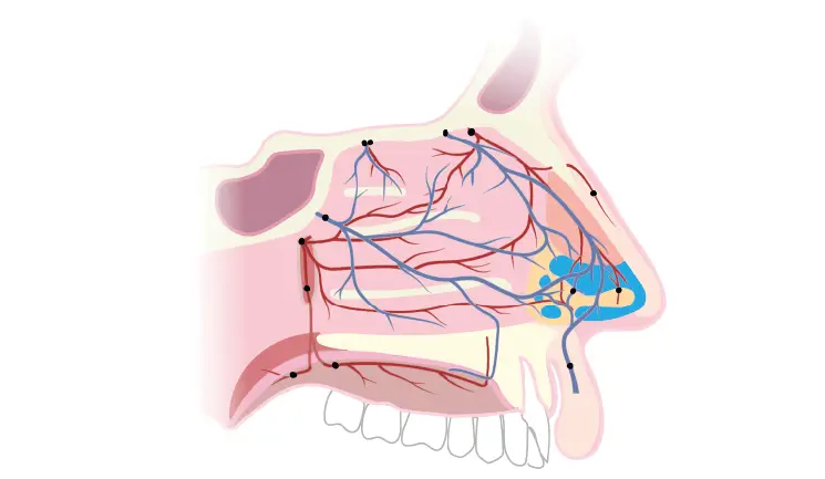
In a case such as this, comprehensive knowledge and understanding of the internal, external anatomy and blood supply to the nose is of paramount importance (Figures 2-4), although it should be noted that the neuro-vasculature may have been compromised and altered due to the previous surgical interventions.2 In addition to this, natural and quite normal anatomical variations often exist. Hence, in order to be able to map all of the essential and intricate anatomy within a fraction of a millimetre, I would strongly recommend the use of a good ultrasound unit, as I did in this case.
There are several key arteries which practitioners need to be aware of when injecting the nose (Figures 2-3). The external blood supply of the nose is derived from key branches from the ophthalmic, infraorbital and facial arteries. There are also additional branches in the form of the external nasal artery, which is derived from the anterior ethmoidal artery, which provides blood supply to the dorsum and is sometimes violated during non-surgical treatment of the nose due to being often overlooked.3
The intricate internal blood supply of the nose should also be considered. It is mainly derived from the third part of the maxillary artery, in the form of the anterior and posterior ethmoidal arteries, the sphenopalatine artery, greater palatine artery and several branches from the superior labial artery (derived from the facial artery). Practitioners must also consider the vascular plexus known as Little’s area or Kiesselbach’s area, found in the anterior nasal mucosa.3
The combination of an in depth knowledge of the anatomy and verification with ultrasound allowed me to plan precise entry points and depth of delivery of the dermal filler, in any given section of the nose. Prior to injection, I also aspirated to help ensure I wasn’t injecting into a significant vessel. Aspiration can deliver false negatives or positives on occasion, but I find it is useful as a guide when selecting my entry points.4 I feel that every precaution taken in cases of this complexity is another step to preserve the safety of the patient.
Figure 3: External blood supply of the nose © Dr Bob Khanna Training Institute. Annotations are as follows: A Supraorbital artery, B Supratrochlear artery, C Dorsal nasal artery, D External nasal branch of anterior ethmoidal artery, E Columellar branch, F Septal branch, G Facial artery, H Superior labial artery, I Angular artery, J Lateral nasal artery, K Infraorbital artery
Figure 4: Osseous-cartilaginous structure of the nose © Dr Bob Khanna Training Institute. Annotations are as follows: A Radix/sellion, B Rhinion, C Dorsum, D Accessory nasal cartilage, E Alar fibrofatty tissue, F Medial crus of major alar cartilage, G Septal cartilage, H Infratip lobule, I Alar margin, J Tip-defining point, K Alar cartilage, L Upper lateral cartilage, M Septal cartilage, N Nasal bone
Due to the patient’s soft tissue scarring from a history of acne and surgery and the thinness of her skin, I had to be very selective about the most appropriate materials to use to achieve a smooth and aesthetically satisfactory result. In this case, I used my own layering technique by selecting two different dermal fillers with varying viscosities: Pluryal Biovolume, which has a higher viscosity, and Pluryal Bioclassic for the less viscous option. I injected Biovolume deep to the nasal superficial musculoaponeurotic system (SMAS) to reinforce the foundations of the nasal structure, and Bioclassic was used superficial to the nasal SMAS to fine tune and create a smoother aesthetic result, without damaging the patient’s delicate skin. I selected these bio-stimulating fillers, to create longer-lasting results through tissue regeneration alongside re-sculpting the nose. Using Matricyl 3Dtechnology, the Pluryal bio fillers facilitate a slow release of free HA and stimulation of mainly CD44 receptors to increase levels of hyaluronic acid and fibronectin, which is essential in neo-collagenesis.5-7
In my view, when encountering a case that has issues like those aforementioned, it is safer to use a cannula, as using needles carries a greater risk of breaching the internal wall of the nose in cases where it has already been compromised.8 I used two different Dermasculpt cannulas: a 22 gauge 70mm for the Biovolume, and a 25 gauge 40mm for the Bioclassic. In this case, I chose to inject the nose via two entry points – one in the nasal tip and one in the mid-dorsum – because access to the nasal tissue was restricted by the contour defects from the patient’s previous procedures. In total, I used just over 1ml of dermal filler to complete this procedure in one session. The patient was extremely happy with the results and felt I had surpassed her expectations (Figure 1).
Aftercare in a case like this is crucial to help preserve the results achieved. I warned my patient not to wear glasses of any kind for two weeks following treatment, not to blow her nose with force and to sleep on her back for a fortnight to reduce the risk of the nose receiving any trauma whatsoever. I also told her to avoid either very hot or very cold showers/baths as the extreme temperatures can affect blood flow and therefore affect the healing process. I discouraged her from using any highly active skincare on her nose for two weeks, instead recommending a moderate-strength glycolic acid on the nose both morning and night, with SPF 50 in the mornings and a gentle moisturiser before bed. All products were from my skincare line, DrBK Skin. I made sure the patient was aware that rare delayed complications could arise after treatment, so informed her to report back to me if she noticed any irregular skin discolouration, swelling or changes in skin texture.9
© Dr Bob Khanna Training Institute
When presented with complex cases like the one described in this article, practitioners must take a step back and assess their own capabilities before deciding whether to proceed with treatment themselves or refer on to a more experienced colleague. So as to safeguard patient safety, an advanced knowledge of anatomy is perhaps the most important factor in order to assist in planning and allow you to fully interpret ultrasound imaging. Having a sound technique with carefully selected products is essential to create a good result and prevent further damage.
Upgrade to become a Full Member to read all of this article.
Aesthetics Media Ltd, 70 London Road, Regal House, Twickenham, TW1 3QS