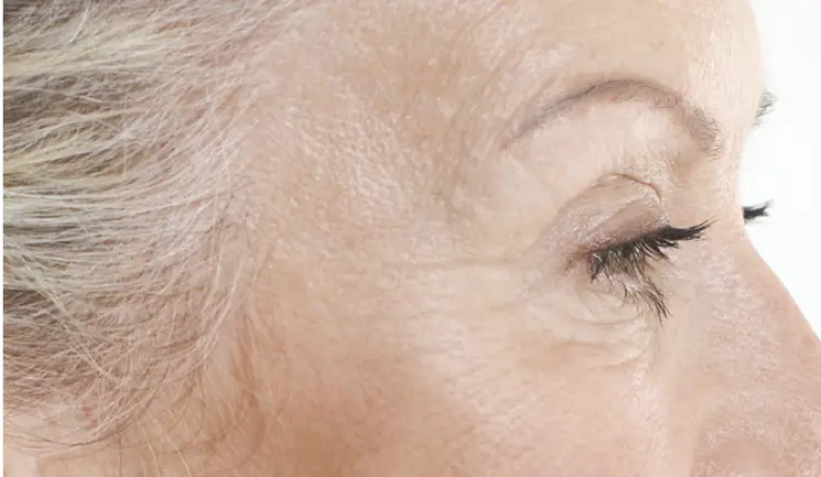
Dr Ciara Abbott details the anatomical considerations of the ageing temple and shares techniques for successful treatment
The temporal region is a frequently overlooked area in facial rejuvenation, yet its impact on the ageing face can be dramatic. Patients are often unaware of the negative impact of hollow temples on their appearance and due to the gradual ageing of this area, rarely request specific temple treatment. However, it is a key area that practitioners should actively assess and consider treating due to its wide range of positive outcomes, including improvement of deep periorbital lines, lifting of the lateral brow and rebalancing of the facial structure to create a more youthful appearance.
In order to treat this important region successfully and without complication, it is essential to understand the anatomy of the temple and how the ageing process impacts the bony and soft tissue dimensions.
Facial ageing is a complex process of bone deposition and resorption, redistribution of facial fat pads and declining skin elasticity.1 Dynamic bony changes result in significant bone atrophy in some areas supporting the temple, such as the inferolateral aspects of the orbital rim and maxilla, which contrasts with bone deposition and expansion in other surrounding structures, such as the zygomatic arch and supraorbital rim.2
In addition to skeletal changes, there is subcutaneous volume loss through the depletion of fat, reduction of elastin and collagen and loss of the temporal fascia, combined with an excess of orbital and temporal skin.3,4 The result is an apparent hollowing of the temporal fossa and what has been described as the ‘peanut-shaped face’ associated with ageing.5
It is best to approach the anatomy of the temple in two ways; the first being to understand the surface anatomical landmarks to demarcate the region itself, which ensures safe injection and helps avoid vascular complication and, secondly, to appreciate the complex soft tissue layers overlying the temple fossa. The four borders of the temporal region are recognised as: superiorly the temporal fusion line, inferiorly the superior border of the zygomatic arch, medially the lateral edge of the orbital rim and laterally the temporal hair line.6,7
The most important of these is the easily palpable temporal fusion line, which allows for reliable location of the ‘safe zone’. This is widely accepted to be 1cm superiorly along the temporal fusion line (caution should be taken if using the tail of the brow as the starting point due to medical and cosmetic alterations of the patient’s original brow), and 1cm inferior to this point (Figure 1).6 Not only is this safe zone most likely to be free of important vasculature (superficial, middle and deep temporal arteries, middle temporal vein) but also is the shallowest part of the temporal fossa to make contact with periosteum and allow for ease of injection, if choosing to administer product with a needle.
Secondly, when considering temple anatomy, it is essential to understand the relationship of the tissue planes and their individual depth. This allows for appreciation of where product should be correctly placed for optimum results.
Figure 2 highlights the multiple soft tissues layers originating from the cranium and their intimate relationship with one another. Although there is some discrepancy amongst anatomists as to the particular correlation these tissues, it is widely accepted that to treat the deep temple safely, and to obtain prime cosmetic outcomes, product needs to be placed ideally on the periosteum and inferior to the temporalis muscle. However, the temporalis muscle is often significantly adhered to the periosteum.
As a result, product deposition here is actually likely to be intramuscular but practitioners should still ensure it is inferior to the deep temporal fascia.8 Within the fascial layers runs significant arterial supply and therefore, again, comprehension of the soft tissue relationship to the periosteum will prevent vascular complications.
As appreciation for rejuvenating the temple has increased, so too has the evolution of product choice. Various treatments are utilised in this area, ranging from autologous fat transfer, to polylactic-L-acid (PLLA) filler and the more commonly-used hyaluronic acid (HA) fillers.
Within my own practice I regularly use HA dermal fillers to treat the temple, for several reasons. Firstly, HA fillers are thought to be more stable and less likely to migrate in this area than alternative fillers mentioned above.10 In addition, they provide immediate cosmetic results, and therefore allow the practitioner to actively assess the appropriate level of rejuvenation required and, thus, prevent overcorrection of this subtle area. Furthermore, their reversibility makes for a sensible choice in this complex anatomical region.
When choosing which HA filler to use, one should consider the thickness of the dermis of the temporal skin, which, along with periorbital skin, is known to reduce in thickness with ageing. It is therefore important to consider filler viscosity to prevent visible skin complications but, in practice, if treating the temporal defect by deep periosteal injection, this should be of less clinical concern.
In my experience, due to the large volume loss often seen in this area, a highly crosslinked, viscous product with a high G prime, is likely to produce better ‘lift’ and will provide potential longevity of treatment for the patient.
Volume of product is another consideration as, in order to achieve visible results, large volumes of HA filler may be required. From experience, in the majority of cases an average of 0.5-1ml of HA filler in each temple is required to rejuvenate this region, which may be undertaken in a sessional approach, and it is essential to have this discussion with your patient prior to treatment.
Temple rejuvenation has been previously approached with both cannula and needle, and the differences in technique and outcomes have been extensively discussed between aesthetic professionals.11 It is generally accepted that use of cannula in this region needs to be with a blunt tip and inserted superficially i.e. interfascial or subdermal; the benefit being reduced vascular trauma and potentially less product requirements.12 However, I find that contour irregularities are a potential complication with superficial placement.
More commonly is the use of needle injection, down to periosteum using a bolus technique.6,12,13 By being down to bone, one can be more confident that they are unlikely to be within a vessel, due to the vasculature in this region running within the soft tissue layers, for example, in the temporalis muscle and above. Herein we discuss my approach to use of needle injection for temple rejuvenation.
It is essential with every individual patient to accurately mark the anatomical landmarks prior to treating the temple region. Once the location of the ‘safe zone’ has been identified, using the 1cm superior and inferior rule, the practitioner needs to palpate the ‘safe zone’ again to ensure there is no palpable pulse. This can be quite subtle and easy to miss; therefore, I would advise to feel for this with an ungloved finger.
Once the clinician can be assured there is no arterial pulse felt, it is important to next look closely at the skin within the ‘safe zone’ to ensure there is no prominent venous supply. A surgical light can highlight this adequately, or asking the patient to lean forward and perform the Valsalva manoeuvre can emphasise any hidden venous supply which can subsequently be avoided.
After ensuring there are no vessels at the site of injection, clean the skin and adopt appropriate aseptic technique. Then, using a 27 gauge needle, with entry perpendicular (90 degrees) to skin, I insert the needle deep to bone, cautiously ‘touching’ the periosteum to avoid trauma to the thin temple bone. This is followed by aspiration to ensure no return of blood in the needle hub.
If right handed, hold the syringe in the right hand, aspirate with the left and then immediately place the index and third finger of the left hand straight onto the patient’s hair line and apply light pressure to this area during the injection. This prevents posterior spread of product. Injection here needs to be extremely slow with even pressure. During this slow delivery of product, the injector should be able to visualise the gradual lift of the temporal hollow, which not only confirms the correct position and placement of the product, but also allows for the injector to continually assess the amount of product required whilst injecting. Of course, if left handed, adopt the opposite approach.
It is important to bear in mind potential asymmetry, with the left and right temple potentially requiring different amounts of product due to uneven volume loss, and also to appreciate the need for under-treating the area. The end result should ensure an acceptable subtle concavity, particularly in females, to prevent masculinisation that occurs with a convexity or overcorrecting of the temple.
As with all dermal filler procedures, treating the temple can result in common side effects such as bruising, due to the thinness of the temple skin and/or swelling. In my experience, this can be counteracted by gentle compression of the injected area. Furthermore, patients can often describe the area as being tender some hours after the procedure, so general aftercare advice and post-treatment analgesia is beneficial. In addition, overcorrection of temple deficit by excessive product injection will result in an unwanted cosmetic outcome, so it is therefore best to always under treat and consider treatments in several sessions to avoid this.
Serious complications have been documented when treating the temples with filler, and therefore anatomical understanding is key. The most significant of these are vascular complications, namely retinal occlusion resulting in iatrogenic blindness, when filler is injected inadvertently into the ocular circulation.14
To avoid these potentially catastrophic complications, I would recommend that the practitioner should adhere to the following:
Treatment of hollow temples is often an under-appreciated part of facial rejuvenation, both by patients who overlook this subtle area of ageing, and sometimes by practitioners who may feel the potential side effect risks outweigh treatment benefit.
In my experience, careful mapping of anatomy, precise marking of vasculature and the ‘safe zone’, and treating the temple with needle down to periosteum, with aspiration and slow injection, is the safest and most reliable way of treating this complex area. Counselling your individual patients about potential risks and side effects versus their possible aesthetic improvement is key to a successful outcome when treating the temple with HA filler.
Upgrade to become a Full Member to read all of this article.
Aesthetics Media Ltd, 70 London Road, Regal House, Twickenham, TW1 3QS