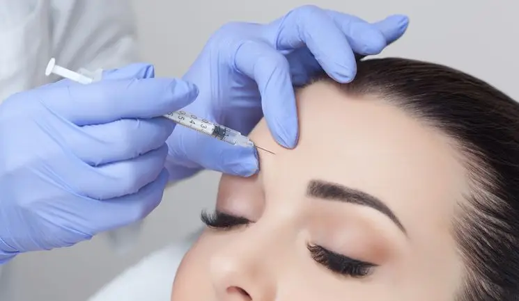
Treating the Glabella with Dermal Filler

By Chloé Gronow / 02 Oct 2020

Practitioners detail the benefits and challenges of using hyaluronic acid in this high-risk area

The glabella: one of the main facial features to display signs of ageing. When rhytids form in this area, they will generally range from fine lines to deep furrows. For many, their presentation does not simply present an aged appearance, but can make individuals look angry, sad, anxious, fatigued or fearful.1 Anecdotally, practitioners explain that this can cause undue distress in patients who feel that their emotional connection with friends, family and colleagues is being negatively affected by their appearance.

Thankfully, we all know treatment options are available. Botulinum toxin A has become a mainstay; inducing temporary paralysis when injected into the striated muscles and, thus, reducing the appearance of rhytids in the glabella.1 The well-established treatment is usually successful and low risk, so why use anything else you may ask?

Well, as aesthetic practitioners Dr Beatriz Molina and Dr Raul Cetto explain, administering hyaluronic acid (HA) dermal filler in the glabella can offer enhanced results for those with significant furrows, while also being longer lasting.2 That said, injecting HA in this area is off-label with some products and comes with higher risk than toxin or the use of HA in other facial locations. This article will explore the research, potential complications and precautions to take if it is a treatment option you’re considering offering your patients.

Practitioner concerns

According to Dr Molina and Dr Cetto, many practitioners became fearful of treating the glabella with filler after the publication ofAvoiding and Treating Blindness from Fillers: A Review of the World Literature in 2015.3 In the paper, 98 cases of vision changes from filler were identified, with the glabella region being the highest risk area, with 38.8% of complications occurring there. Autologous fat was the most common filler type to cause the complication in all cases (47.9%), followed by HA at 23.5%.3

An update to the research was published in 2019, which identified 48 new reported cases of partial or complete vision loss after filler injection between January 2015 and September 2018. This time, the glabella was the second highest risk area (27.1%), following the nasal region where 56.3% of the complications occurred. HA filler was the cause of complications in 81.3% of the cases.4 Dr Molina and Dr Cetto suggest that complications involving the glabella may have gone down because, anecdotally, less practitioners have been injecting the area with HA since the 2015 report. Dr Cetto says, “Five years ago it became almost like a taboo to treat the glabella with filler, yet many practitioners had been treating the area routinely before then and achieving successful results.” Dr Molina explains, “When I started 15 years ago, you were taught to treat the glabella from the beginning of your training. Since we’ve become more aware of the anatomy and potential complications, however, there’s been a big shift to practitioners refusing to treat the area.”

While they both acknowledge the serious risks associated with treating the glabella with HA, Dr Molina and Dr Cetto emphasise that it can be safely and successfully treated by experienced practitioners – not beginners – who have an excellent understanding of the anatomy and are appropriately trained. Doing so, they believe, will offer an overall enhanced result of the whole area.

Assessment and consultation

Dr Molina and Dr Cetto agree that using HA in the glabella should always be the secondline treatment, following the administration of toxin. “I don’t think there’s a shortcut,” says Dr Cetto, explaining, “If you just administered filler, you won’t take away the core component causing the concern, which is the muscle contraction deepening the groove or line.” Ultimately, he says, the patient won’t be satisfied with the result.

The pair note that they would usually consider HA treatment in patients with deep static lines. “You’ll realise that sometimes botulinum toxin is not enough to create the significant effect sought by patients, so adding filler afterwards is of real benefit,” says Dr Molina, explaining, “It can help if there is significant brow ptosis and volume loss in the area.”

Dr Cetto adds, “As we know, muscles become hypertrophic from frowning a lot and skin loses its elasticity. When a patient has a static line with a dip or a groove it is very often not that easy to treat with toxin alone.” He continues, “Changing a patient’s expression through treatment can really help. Of course you’ve got to evaluate whether filler is appropriate on a case-bycase basis for every person you see.”

Dr Molina will follow-up with potential patients approximately two weeks after their toxin treatments to assess the result and decide whether HA will benefit. “As the toxin will have taken effect, you’ll have a good understanding of the result and can outline the next steps to the patient. Some people will prefer to wait a few months; it just depends on the severity and the amount of perfection the patient is looking for.”

Dr Cetto, on the other hand, prefers to wait to review after two cycles of toxin to decide whether filler would benefit the patient. The only time the practitioners would use HA alone in the glabella is if the patient specifically does not want toxin treatment or if they have a contraindication. Dr Cetto exemplifies a case of a patient who is a teacher who did not want toxin injections; while she wanted to soften the deep line in her glabella, he explains she did not want to relax the muscles in the area as being able to frown was an important part of her role when engaging with her pupils.

Both practitioners emphasise the importance of detailing the risks of the treatment to the patient, both verbally and in their written consent form prior to treatment. “It’s not about scaring the patient, but we should make them aware that there’s higher risk when injecting the glabella with HA compared to, for example, injecting the lip,” says Dr Cetto, adding, “We should make sure they’re fully aware of what could go wrong, how we will manage it if the worst happens and, like with any treatment, ensure they are given an appropriate cooling-off period of at least two weeks to consider whether they definitely want to go ahead.”

Anatomy

The practitioners interviewed emphasise that the first thing to remember when injecting the glabella is that not everyone’s anatomy is the same. Dr Cetto notes that both the location and the depth of blood vessels can vary between patient so there’s no guarantee of safe areas to inject. Dr Molina and Dr Cetto explain that the key arteries to be aware of are the supratrochlear and the supraorbital arteries (Figure 1), which are both branches of the ophthalmic artery. As detailed in the peer-reviewed article Anatomy, Head and Neck, Glabella, these arteries supply the forehead and medial canthal area. The supraorbital artery passes through the supraorbital notch, where it divides into two branches: a superficial branch and a deep branch. Superficial branches include the vertical and brow branches, while the medial, oblique, and lateral rim branches are deep.5 The article explains that the supraorbital and supratrochlear arteries anastomose with the angular artery at the medial angle of the eye to form an arterial arcade. The supratrochlear artery also branches from the ophthalmic artery and exits the superomedial orbit.

The authors claim that the artery becomes subcutaneous 15 to 25mm above the supraorbital rim; it can be relatively superficial, being about 2mm deep in the muscle layer. A branch of the dorsal nasal artery supplies the glabella and the inferior and middle transverse regions of the forehead; laterally it anastomosis with supratrochlear arteries.

In a 2019 article called Forehead Anatomy for Injectables, nurse prescriber Anna Baker and consultant plastic and aesthetic surgeon Mr Dalvi Humzah highlight a study that has suggested that the deep vertical lines observed in individuals may serve as a marker for the underlying supratrochlear arteries in 50% of cases.6 The artery is, however, variable in its arrangement.

According to the authors, the superficial branch is always present, but the deep branch is not present in more than 80% of cases. This has been used as a basis of dividing the arterial patterns of the forehead into two types: type I – with superficial branches of the supratrochlear and supraorbital artery, with the deep branch of the supraorbital artery – and type II – with both superficial and deep branches of the supratrochlear and supraorbital artery.

Baker and Mr Humzah explain that the types are further divided depending on whether a central dorsal artery is present medial to the superficial branch of the supratrochlear artery. They note that this variability should be considered when dermal filler injections are being performed in the forehead, and deep placement with a cannula is recommended. The authors continue that the supraorbital and supratrochlear arteries travel with corresponding nerves and the supraorbital branch exits the medial aspect of the orbital rim along its superior course to the frontalis. An accessory foramen may be located just superior to the supraorbital foramen, which is a possible and significant anatomical variation to note when assessing an individual’s glabellar complex.

Whilst it is acknowledged that the glabella is considered a higher risk area owing to the medial and variable anastomosis of the supratrochlear vessels, Baker and Mr Humzah remind practitioners that they are advised to remain mindful of the extensive and varied anastomosis of the entire facial vasculature.

Product selection and technique

Dr Molina and Dr Cetto highlight that a HA filler with a moderate G-prime is best for treating this area. Dr Cetto says, “You want a product that’s cohesive but not stiff,” while Dr Molina adds, “It needs to be soft enough to integrate well and look natural, as most people don’t have much fat in their glabella so a thicker product can make it look lumpy. You also want it to support dynamic movement, so the patient can still have brow expressions.”

Teosyal RHA 2 is Dr Cetto’s product of choice, unless he is injecting a patient with particularly thin skin – he would then use RHA 1. “RHA 1 is less strong and more stretchable,” he explains. He usually injects the glabella into the mid-dermis with a 27 gauge needle, at a 10 degree angle.

“I go perpendicular to the line, rather than going along the line in a retrograde fashion,” Dr Cetto explains, highlighting, “We know the arteries are more likely to be along the line, so you could be more likely to injure them by injecting this way.” Dr Molina uses a cannula in this area, explaining that there are two possibilities to make an entry point, which she usually does with a 23 gauge needle. “You can identify the middle point in-between the supratrochlear and supraorbital nerves or, if you’re a bit nervous of that small space, you can enter more laterally on the brow. Look at where the middle point of the middle and lateral brow meet and you can enter from there,” she explains.

Dr Molina then switches to a short 25mm 25 gauge cannula which, she notes, offers good control. “If you’ve gone in laterally through the brow there will be a slight bending, so you should guide the cannula with care. Know your layers to ensure you’re above the blood vessels when you place the cannula. Be slow and gentle; don’t push hard as you could move vessels,” she emphasises.

For Dr Molina, using a product without lidocaine is important. “As pain is one of the most common identifying factors of a vascular event, when injecting the glabella I want patients to be able to tell me if they are experiencing pain,” she explains, highlighting, “The problem with lidocaine is the area goes numb after the first injection, so it can be very nerve-wracking as, without the patient reacting, you may not be not aware that you’re applying pressure on the blood vessels.” As such, Dr Molina uses Aliaxin FL, which she explains is a soft filler that integrates well in the glabella and gives a very natural result.7

Risk management

Dr Molina explains that bruising is the most common side effect to occur when treating the glabella with HA. “You may hit a blood vessel superficially, so a bit of tenderness is expected,” she explains.

But of course, the most serious risk of injecting the glabella with HA is vascular occlusion, potentially leading to skin necrosis or blindness. Prevention is always better than cure, so having a thorough understanding of the anatomy, injecting slowly and with care, and using small amounts of product is essential to avoid such complications, note the practitioners, as well as the authors of the 2015 literature review.3

Dr Molina recommends aspirating on each injection if using a needle and stopping immediately if the patient experiences pain or a change of colour or blanching is noted. If this occurs, then practitioners are advised to implement their vascular occlusion management strategy straightaway. The International Association for Prevention of Complications in Aesthetic Medicine (IAPCAM)8 and the Aesthetic Complications Expert (ACE) Group9 provide valuable guidance on this, highlighting the importance of having an emergency kit in reaching distance. Dr Cetto comments that aspiration is not always a reliable method of assessing if you are intravascular, noting that one recent consensus paper on preventing blindness does not recommend it.10

For Dr Molina, her approach involves always having a vial of hyaluronidase and 1ml of sodium chlorate next to her, so she is ready to mix and inject the solution immediately. The volume can be increased once you have assessed and noted what is needed for the particular scenario, she advises. Speaking from experience of managing a vascular occlusion immediately after injection in the nose, Dr Molina highlights that in such an emergency situation there will be multiple things to do, all while needing to remain calm and reassuring the patient at the same time.

“Having everything prepared in advance will make the process more manageable and ease any anxiety experienced by the patient or yourself,” she says, noting it can also reduce the possibility of long-term damage.

To treat or not to treat?

You may currently treat the glabella with HA confidently, but are you fully aware of the risks? Or maybe it’s something you’ve thought of doing after hearing success stories, but not known how best to approach?

This article has aimed to inform you of the risks and considerations to bear in mind and help you make an informed decision. It is not an exhaustive list and practitioners are always advised to speak to their peers and complete appropriate training, both in injecting the area and in managing complications, before attempting any new treatment on patients.

As highlighted, treating the glabella with HA is considered high risk so if you decide to go ahead, Dr Molina and Dr Cetto advise to be thoroughly confident in facial anatomy, undergo extensive training and ensure you have the qualifications and knowledge of exactly what to do if a complication does occur. They also recommend checking with your insurer that you will be covered to perform the treatment off-label if the products you use don’t have instructions for use. If treating the glabella is not for you, the practitioners highlight the value of having a network of colleagues who you can refer to. Dr Cetto concludes, “In my experience, patients appreciate your honesty and, if you do refer, their trust in you will increase, meaning they will always return to you for their usual treatments.”

Upgrade to become a Full Member to read all of this article.