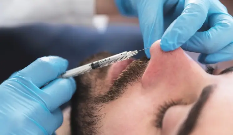
There has been a decline in the number of people undergoing surgical rhinoplasties.1 At the same time, an increasing number of patients are undergoing non-surgical rhinoplasty (NSR) annually.2 Although appearing to be non-invasive, the NSR comes with a significant risk. This article will outline relevant nose anatomy, and the valuable role of ultrasound in the prevention and management of vascular complications in the nose.
A comprehensive understanding of relevant anatomy is key to a safe non-surgical augmentation using filler.
The nose usually consists of five layers:3
The arteries of the nose encountered during NSR originate from both the internal carotid (via the ophthalmic artery) and external carotid (via the facial artery) with various anastomoses between the two systems.4 However, below is a basic account of the common arteries that can be encountered during NSR:5
The vascular supply of the nose is usually found superficial to the musculature.