With society’s levels of stress and anxiety increasing, the number of stress-related teeth clenching and grinding cases are on the rise.1 However, these symptoms are just clinical manifestations of a more complex problem called temporomandibular disorders (TMDs).2 They are often accompanied by enlargement or hypertrophy of the masseter muscle, giving the lower third of the face a squared appearance. This is an unwanted aesthetic appearance for many female patients, leading to an increased demand for an aesthetic treatment which can aid facial slimming.3
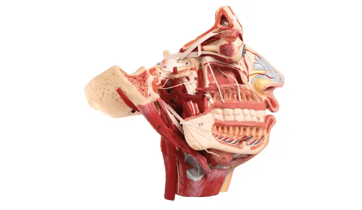
When administering botulinum toxin for facial slimming or treating temporomandibular disorders, knowledge of anatomy and topographic anatomy of the area is paramount. This article will describe the anatomy and function of the muscles of mastication, as well as the anatomical relations of them.
Aesthetically undesired hypertrophy of the masseter muscle is often caused by temporomandibular disorders which are one of the leading causes of orofacial pain.1 They involve dysfunction of the…